|
What
is nearsightedness ?
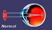
Nearsightedness
(myopia) is when the eye is too long or the curvature
of the cornea is too steep and the focus of the
rays of light that enter the eye fall short of
the retina. The result, is a blurry view of distant
objects.
|
|
|
What
is astigmatism ?
Astigmatism
can exist alone or in combination with nearsightedness
or farsightedness. With this condition the cornea
becomes oval-shaped like a football instead of
round, causing distortion when the eye tries to
focus.
|
|
|
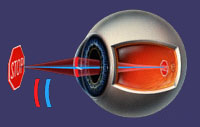
What
is farsightedness ?
Farsightedness
(hyperopia) occurs when an eye is too short or
the curvature of the cornea is flat. Light rays
entering the eye focus behind the retina, and
as a result a blurred image is produced, especially
with near objects.
|
|
|
What
is presbyopia ?
Presbyopia
is when the lens of the eye looses the ability
to change focus. This occurs as part of the natural
aging process and usually begins around 40 to
45 years of age. When we lose this ability to
change focus it prevents us from seeing both near
and distance simultaneously. A person will need
to have extra magnification in his or her glasses
in the bottom or bifocal part. A nearsighted person
over 40 or 45 years of age will be able to see
up close if they remove their glasses since their
eyes naturally focus at near, but if they wear
distance glasses they will then need a bifocal
if they want to see near. Since this is part of
the natural aging process, a person will develop
the lens changes whether they have refractive
surgery or not. |
|
|
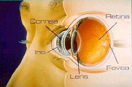
Anatomy
of the Eye
Cornea :
The
cornea is the clear front surface of the eye.
This clear tissue is like a window for the eye.
It is composed of 5 different layers. The outer
layer is called the Epithelium then comes the
Bowman's Membrane, then the Stroma, then Descemet's
membrane, the fifth layer is called the Endothelium
and is on the inside of the eye. The cells and
fibers in the cornea are arranged so that light
can pass through it with a minimum of diffraction
and internal reflection. The cornea contains no
opaque substances such as blood vessels that would
mar it's clarity. It receives its nourishment
from the vessels surrounding the cornea. It is
kept shiny and lubricated by tears that keep its
surface moist. |
 |